
What is an atypical mole? - Sporadic dysplastic moles and familial atypical mole syndrome explained.
On Jun 18, 2013
-
Keypoint Tweet @CentralSkin: The ugly duckling mole is no match for the ABCDE criteria!
-
Moles are made up of melanocytes
Moles, also known as nevi, are very common growths of the pigmenting cells in your skin. The pigment cells that grow are called melanocytes. The density of melanocytes in your skin determines the lightness/darkness of your skin tone. Melanocytes also protect the DNA of your skin cells from the sun’s harmful UV rays. Moles can either be congenital or acquired during childhood. Moles can become atypical and can even develop into melanoma, a skin cancer formed by abnormal proliferation of your melanocytes.
-
Dysplastic nevi are moles that possess atypical characteristics
Atypical moles, also called dysplastic nevi, have certain defining characteristics that may raise concern for melanoma and may require a biopsy. However, if you have been told that you have an atypical mole, you should not think that this means that you are destined to develop melanoma sometime in the future. There are many individuals who have one or several atypical moles and whose risk for melanoma is not much higher than individuals without atypical moles. Nonetheless, careful follow-up with your dermatologist is required. Atypical nevi are most often seen in Caucasians, especially those with light hair and light eyes. Darker skinned populations, including blacks, Asians, or Middle Easterners, rarely develop atypical moles and have a very low rate of skin cancer.
-
Dermatologists use the ABCDE criteria to categorize a mole as atypical
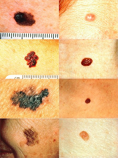
A: Asymmetry. If you can theoretically cut a line through the mole and it could fold over on itself, it is symmetrical. The more asymmetrical it is, the less overlap you would have if you theoretically fold a mole over on itself on a dividing line.
B: Irregular Borders. Moles with jagged or non-uniform borders fit this criterion.
C: Color Variegation. Most normal moles should be fairly uniform in color. However, when a mole has multiple different colors inside of it, such as black, tan, red, brown, or blue, it should raise some suspicion. (Knowledge tidbit: variegation means variation in color)
D: Diameter. Moles that are larger than 6mm or the size of a pencil eraser fit the D criterion. However, you should know that melanoma can present itself as an atypical mole that is smaller than 6mm. Thus, the diameter less than 6mm is not an absolute rule-out factor for melanoma.
E: Evolution. This factor refers to the change in the quality of moles. Benign moles should usually keep their ABCD criteria fairly steady. However, atypical moles may develop and grow larger, attain more irregular borders, show different colors and become less symmetrical. When this occurs, one always has to consider the option that a malignant melanoma may be forming.
-
How one should interpret these criteria?
One must not look at any one of these criteria in an isolated fashion but should instead consider what the combination of all the ABCDE criteria may reveal about a mole. Lack of any of these ABCDE criteria should not be used to exclude the possibility of melanoma.
-
What is the “ugly duckling” mole phenomenon?
People usually have a pattern or "signature" in which they form most of their moles. Spotting an “ugly duckling” mole that does not look like the rest should raise suspicion for an atypical mole or melanoma.
-
What is Familial Atypical Mole Syndrome?
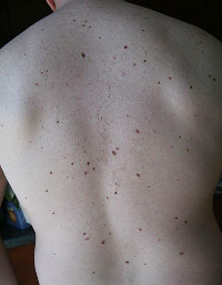
It is a genetic condition in which patients have many atypical moles and have a very high risk of developing malignant melanoma. These patients may typically have either first-degree (mother, father, siblings) or second-degree (grandparents, uncles, aunts) relatives who have a history of melanoma. These patients have many moles, oftentimes more than 50. While some of these moles are benign, some may be atypical and require excision. Upon excision, the moles may show dysplastic, or atypical, features when examined under a microscope. Familial Atypical Mole Syndrome is inherited in an autosomal dominant fashion, meaning that a child has 50/50 chance of developing the Syndrome if one parent has it and has 100% chance of developing the Syndrome if both parents have it.
-
Bottom line:
All individuals should have a full body skin exam by a board certified dermatologist. If your dermatologist detects an atypical mole, they may elect to perform a biopsy of it. However, if they strongly suspect melanoma, they will skip the biopsy and perform a complete excision of the mole. If you have had multiple biopsy-proven atypical moles diagnosed in the past, you have to take extra precautions to avoid excessive sun-exposure, always wear a broad-spectrum sunscreen with at least an SPF of 30. In addition, you should perform self skin exams every 2-3 months and follow-up with your dermatologist as recommended. If you have been diagnosed with Familial Atypical Mole Syndrome, you should have consistent follow-up with your dermatologist, be very diligent about sun exposure, and perform frequent self full skin exams. Your dermatologist may even recommend that you undergo full-body photography that records the position and visual characteristics of every mole in your body. You and your dermatologist will use these photographs to compare any changes in your moles in the future. Being proactive and vigilant will give you and your dermatologist the best chance of detecting melanoma in its early curable stages.
Comments
Leave a Comment